Kuo-Cheng Lo, Yung-Chun Hsieh. (2016) Comparison of Ball-And-Racket Impact Force in Two-Handed Backhand Stroke Stances for Different-Skill-Level Tennis Players. Journal of Sports Science and Medicine(15), 301 - 307.
Kuo-Cheng Lo, Yung-Chun Hsieh. (2016) Comparison of Ball-And-Racket Impact Force in Two-Handed Backhand Stroke Stances for Different-Skill-Level Tennis Players. Journal of Sports Science and Medicine(15), 301 - 307.
This study compared the kinetic roles of the upper extremities in racket impact force generation between the open stance (OS) and square stance (SS) for tennis players with different skill levels in two-handed backhand strokes. Twelve male tennis players were divided into an advanced group (AG) (L3-L2 skill level) and intermediate group (IG) (L7-L6 skill level), and their data were used in a three-dimensional kinetic analysis. Their motions were captured using 21 reflective markers attached to anatomic landmarks for two-handed backhand stroke motion data collection. During the acceleration phase, significant differences were not observed between both stances, but they were observed between the groups with different skill levels for the force of the upper extremities (p = 0.027). The joint forces were significantly lower in the AG than in the IG. Players performing the SS had significantly larger pronation and supination of the wrist joint moment than those in the OS (p = 0.032) during the acceleration phase, irrespective of the playing level. Higher internal rotation moment after impact was observed at each joint, particularly among young intermediate tennis players, regardless of their stance. The AG demonstrated a higher joint force and moment at every joint compared with the IG at impact. Moreover, the AG demonstrated superior stroke efficiency and effectively reduced joint moment after impact and sports injury.
Advanced players, regardless of open stance or square stance, have larger joint force and moment at each joint before ball impact resulting in better stroke efficiency and reduced chance of injury.
Intermediate players, regardless of stance, have higher internal rotation moment at each joint instead of larger joint force as compared to advanced players before ball impact. The higher internal rotation moment will induce higher joint impact force which makes the player injury-prone.
Young intermediate tennis players may want to avoid excessive follow-through movement after ball impact to prevent injury in their early career.
INTRODUCTION
Because tennis is a racket sport, effective integration of the entire body segments is essential for transferring the ground reaction force to the racket through the trunk and upper extremity. Repeat impact from the ball hitting the racket when one-handed backhand stroke is performed is believed to be related to tennis elbow syndrome (Roetert and Groppel, 2001). Moreover, typical tennis activities require an intact kinetic chain to create energy and transfer the force through the joints. Disruption of the kinetic chain can increase the loading on the other joints in the sequence of movements and cause potential injury (Kibler, 1995; Kibler and Sciascia, 2004; Marshall and Elliott, 2000).
Studies on backhand stroke mechanics have compared elbow muscle activities in one- and two-handed backhand strokes (Giangarra, et al., 1993) and quantified joint angular movement differences between the two stroke styles (Akutagawa and Kojima, 2005; Genevois, et al., 2015; Kawasaki, et al., 2005; Reid, 2002). The joint loading on the smaller lower lumbar spine and hip joint in the one- and two-handed backhand strokes as well as the momentum transfer during a two-handed backhand stroke (Stępień, et al., 2011; Wang, et al., 2010) can be predicted through an inverse dynamic analysis of foot loading and kinematic data (Akutagawa and Kojima, 2005; Kawasaki, et al., 2005). Although body movement and joint loading information are provided through these kinematic, kinetic, and kinesiology data, the energy or force transfer during a stroke still cannot be identified. Because of the fast tempo of modern tennis competitions, the open stance (OS) stroke is more widely adopted (Wang, et al., 2010). Previous studies have compared the differences between the OS and square stance (SS) in two-handed backhand strokes; however, only Wang (2010) focused on the momentum transfer from the racket to the ball during the ball–racket contact. Regardless of the skill level of players, significantly higher backward linear momentum contribution (p < 0.05) in the trunk and upper arm was observed in the SS and higher shoulder rotational angular momentum was observed in the OS. The external rotators in the upper arm generate the power in a backhand stroke. The two-handed backhand stroke is powered not only by the trunk rotation, but also by the same amount of angular momentum generated at the shoulder and wrist.
This study compared the kinetic roles of the upper extremities in the racket impact force generation between the OS and the SS for different-skill-level tennis players in two-handed backhand strokes. We hypothesized that the stroke stance and skill-level differences exist in the measures of the impact force, advanced group (AG) demonstrates more efficient force generation than the intermediate group (IG), and SS is responsible for more efficient force transfer. This knowledge of the biomechanics of the upper-limb joints will be helpful in improving the efficiency of training protocol designs and can assist physiotherapists in rehabilitation planning.
METHODS
Twelve male tennis players participated in this study after providing written informed consent. The players were divided into an AG (Age: 24.2 ± 2.3 yrs; Height: 1.72 ± 0.04 m; Weight: 67.3 ± 6.7kg; Experience: 9.7 ± 2.4 yrs; L3-L2 skill level) and IG (Age: 15.2 ± 1.0 yrs; Height: 1.73 ± 0.04 m; Weight: 58.3 ± 2.9 kg; Experience: 2.9 ± 1.4 yrs; L7-L6 skill level) according to the International Tennis Number Rating System. This study was approved by the National Cheng Kung University Hospital Human Experiment and Ethics Department (ER-95-121), and all participants signed committee-approved informed consent forms.
The two-handed backhand stroke motion was captured using an eight-camera Eagle® motion system (Motion Analysis Corp., Santa Rosa, CA, USA) at a sampling rate of 500 Hz in an indoor tennis court. The tennis ball machine (Tournament model 401, Lobster Inc., Plainfield, NJ, USA) was positioned 12 m from the tennis court baseline, and the ball was served at a speed of 14 m/s. The same tennis racket (Prince precision spectrum 670, string tension is 249 N) was used for stroke motion collection. The racket mass was 0.380 kg, the racket length was 0.686 m, and the center of the racket mass location was 0.344 m from the bottom.
The coordinate system of the trunk, upper arm, forearm, hand, and racket was defined using 21 reflective markers attached to the racket and subject. These markers were attached to the xiphoid process, sternal notch, spinous process of the seventh cervical and eighth thoracic vertebrae, left and right acromioclavicular joints, medial and lateral epicondyles of the elbow, radial and ulnar styloid processes, knuckles II and V, anterior superior iliac spine, posterior superior iliac spine, and a triad of markers on the upper arm. The relationship between the epicondyle and triad markers on the upper arm defined in the anatomical neutral position was used to determine the medial and lateral epicondyle marker positions during motion. The ball speed after the ball–racket contact was detected using retroreflective tape wrapped around the ball. In the testing environment, the subjects were allowed to practice the two backhand stances. As the motion collection commenced, the subjects stood on the left side of the tennis court baseline to execute the backhand trials. Each session involved a 1-s static anatomical neutral position followed by ten successful strokes with a 3-min break between the two stroke stances. A stroke was considered successful only when the ball was hit into a specific 2 × 2 m area located in the right corner of the opposite baseline. An average of ten successful trials in each stroke stance was required for this test.
A five-segment linkage system, including the trunk, upper arm, forearm, hand, and racket, was modeled in this test. For the spatial kinematic description, each segment was considered a rigid body and each joint was assumed to be of the ball and socket type. The anthropometric database by McConville et al. (1980) was adopted. The racket weight and center of mass position were quantified using a weight scale and suspension method (Brody, 1985). A three-dimensional trajectory of the reflective markers was smoothed, and the derivatives were calculated using a generalized cross-validation quintic spline smoothing routine (Woltring, 1986) at a cutoff frequency of 7.12 Hz.
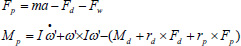
Shoulder, elbow, and wrist loads were defined through the inverse dynamic approach. The complete Newton–Euler equation of motion of each rigid body is given as:
Fp: proximal joint force
Fd: distal joint force
Fw: gravity
Mp: proximal joint moment
Md: distal joint moment
rp: proximal arm
rd: distal arm
ω’: local angular velocity of the body segment
Through calculation of the known distal joint force and joint moment, the proximal joint force and joint moment can be obtained; further, the force and moment of each joint can be obtained.
All force and moment components of the segments in this study were transferred from the segmental coordinate system to the tennis court coordinate system to clarify the segmental contribution in tennis stroke performance. The stroke leading direction, in the court coordinate system, was considered the negative X-axis, referring to the rotation axis of the trunk left–right bending, abduction–adduction of the shoulder and elbow joints, and radial–ulnar deviation of the wrist joint at the neutral position. The Y-axis was the right side of the court, referring to the rotation axis of the extension–flexion of the trunk and body segments. The Z-axis, the upward direction, referred to the rotation axis of internal–external rotation of the trunk and other segments.
The data from the backswing phase (the initial position to the maximum trunk left rotation) to the acceleration phase (the maximum trunk left rotation to the ball–racket contact) and the follow-through phase (from the ball–racket contact to the highest position of the racket) were analyzed. At each phase during backstroke, the skill level and stroke stance effect on impact force generation were analyzed through -a two-way analysis of variance with repeated measures at a significance level of 0.05. The parameter changes during each phase of a stroke cycle were defined as data variations.
RESULTS
Table 1 lists the peak value of the joint impact force and its variation during acceleration. The upper arm, forearm, and hand were in the order of the upper limb mass; therefore, a higher impact force was generated at the shoulder joint during acceleration (F = ma). Table 1 indicates a gradual decrease in the impact force from the proximal shoulder joint to the distal wrist joint. Joint moment varies with impact force during acceleration. Each peak joint moment and its variation are listed in Table 2; the peak joint moments at the shoulder, elbow, and wrist joints occurred in sequence (Figure 2).
During the acceleration phase, the shoulder impact force of the two-handed backhand mainly moved backward and leftward (Figure 1 (a) and (b)). The backward impact force peaked at early acceleration and then weakened immediately. The upward impact force began to decline before the impact, and its variation suggested no significant difference between levels or stances (Figure 1(c)). The shoulder joint’s peak upward impact force was the highest among the other upper extremity joints. During the initial acceleration phase, the shoulder joint moment was observed in major flexion, adduction, and external rotation (Table 2). Abduction occurred at impact and continued to increase with the internal rotation moment after impact. Neither levels nor stances had a significant influence on the peak joint moment and its variation (Table 2).
DISCUSSION
This study compared the kinetic roles of the upper extremities in the racket impact force generation in two-handed backhand strokes between the OS and SS among tennis players with different skill levels. In this section, we discuss the variation in the upper limb joint impact force and joint moment during the acceleration phase when the two-handed backhand was performed in the OS and SS, as well as the joint force and joint moment at impact. Figure 1 and Figure 2 show all the joint impact forces and moments; the values at the moment of impact were excluded to simplify the analysis. This exclusion did not affect the analysis. Only the impact phase data were analyzed, and the coordinate system of the segment was used to describe the direction of the impact force.
Acceleration phase
The higher impact force gradually decreased from the proximal shoulder joint to the distal wrist joint. The impact force variation during acceleration showed that the upper wrist joint impact force of the IG was significantly higher than that of the AG (F1, 10 = 6.684, p = 0.027). A significant difference in the vertical wrist joint (upper/downward) impact force indicated that, compared with the AG, the IG generated higher upward impact force at the wrist joint during the acceleration phase. To avoid backhand stroke failure, tennis players tend to develop an upward spin impact force to raise the racket; therefore, the upward impact force at each joint is higher (Roetert and Groppel, 2001). The joint moment varies with the impact force during acceleration. Each peak joint moment and its variation are presented in Table 2; the peak joint moments at the shoulder, elbow, and wrist joints occurred in sequence and are shown in Figure 2. A significant difference in moment variation was observed only in the wrist joint pronation/supination moment between the stances (Table 2). Neither skill level nor stance caused differences in another joint moment.
The internal rotation moment, which constantly swelled from preimpact to follow-through, exerted a heavy load on the rotator cuff (Figure 2 (c)), and this was particularly true for the IG. This suggests that, to decelerate, intermediate players swing with considerable effort in the follow-through phase, leading to a continual increase in the joint moment in this phase. In addition, studies on shoulder strength and injury have shown that people generally demonstrate greater joint supination strength than pronation strength, which is particularly true of athletes in throwing or racket sports. No difference was observed in the application of shoulder joint strength, regardless of whether the backhand was performed with a single hand or both hands. Shoulder joint injury is most often observed in young tennis players; this is believed to be caused by overuse of the rear rotator cuff (Silva et al., 2006; Bylak and Hutchinson, 1998). Young intermediate players should enhance their follow-through movement after impact and avoid decelerating with considerable strength to minimize the shoulder joint internal rotation moment and avoid injury.
The elbow joint’s force direction during acceleration was in line with that of the shoulder joint. The elbow joint’s rightward and forward forces peaked at midacceleration and then began to decrease. The peak–decelerate phenomenon at the elbow joint occurred later than that at the shoulder joint, indicating that the elbow joint’s impact force was transferred through the limbs. The upward impact force continued to increase until preimpact, and its peak value and variation, as shown in Figure 1 (c), showed no significant differences in the three directions. According to a study by Riek et al. (1999) on the one-handed backhand technique, the forearm muscles continue to move substantially after the impact, and the rightward impact force peaks in the follow-through phase and is then decelerated by the antagonist muscle. Therefore, the horizontal force peaks during the follow-through phase and then declines swiftly; moreover, a leftward force is believed to be generated by the joint impact force moving rightward, which then decelerates.
Elbow joint abduction occurred at early acceleration, at which point the external rotation moment of the elbow joint peaked. The peak external rotation moment during acceleration was 4.78 ± 1.40 Nm for the IG in the OS and 6.71 ± 1.84 Nm for the AG in the SS. After acceleration, the elbow joint adduction occurred immediately and rotated internally until postimpact. The peak value of the internal rotation moment was reached in the follow-through phase, which was attributed to the higher elbow joint impact force after impact. The doubled-handed backhand generated a higher internal rotation moment than did the one-handed backhand, which, according to Kelley et al. (1994), is because the force transferred to the forearm after impact is evenly distributed in the entire elbow in the two-handed backhand but absorbed by the extensor in the one-handed backhand. The two-handed backhand, therefore, generated a lower joint moment than did the one-handed backhand, and this partly explains why tennis elbow occurs more easily when performing a one-handed backhand. Moreover, compared with a one-handed backhand, a two-handed backhand causes fewer sports injuries to the elbow joint (Groppel, 1992).
In addition to the extensor and flexor as the main muscles used at the wrist joint when performing a backhand, Ellenbecker et al. (2006) demonstrated the importance of forearm pronation strength and of pronation strength being greater than supination strength (p < 0.01) in the dominant arm. This is believed to be the result of sports adaptation. Figure 1 shows that the impact force at the wrist joint varied significantly in the acceleration phase. During the initial acceleration stage, because of the two-handed style, the joint impact force was not applied right forward. The wrist joint impact force did not start to move upward and rightward until midacceleration and preimpact. The upward impact force, similar to the shoulder and elbow impact force, continued to increase after impact. According to Figure 1, at this moment, the hands were holding the racket and were ready for impact. After impact, the racket was raised high. The peak values and variation in the forward/backward and rightward/leftward impact forces were relatively close between levels and stances. The IG showed higher upward impact force than the AG (Table 1). This corresponds with the previous statement on sports adaptation and shows the irrelevance of different stances. In addition, electromyography conducted on the one-handed backhand of an AG and IG by Wei et al. (2006) indicated that experienced tennis players are significantly more effective at containing the force and vibration at impact (p < 0.05). Therefore, tiny injuries caused by the forearm muscles at impact, force transmission, and the possibility of the occurrence of tennis elbow can be reduced. The AG demonstrated lower wrist joint force during the acceleration phase. This corresponds with the preceding statement that force transmission is reduced, and the variation in the shoulder and elbow joint force is not significant.
All players demonstrated significantly higher variation in the pronation/supination of the wrist joint moment in the SS than in the OS (p = 0.032) during the acceleration phase, irrespective of the playing level. According to the figures of each joint’s impact force and joint moment during the acceleration phase, the wrist joint was the only joint demonstrating significant differences in the upward impact force between different levels (IG > AG) and in the joint moment between stances (SS > OS). The shoulder joint’s peak impact force and joint moment were the highest, whereas those of the wrist joint were the lowest. The IG’s upward impact force at the elbow joint during acceleration in both stances showed higher peak values than that of the AG (Table 1). This was associated with the IG’s higher internal rotation moment at the elbow joint after impact (Figure 2). Therefore, this was attributed to experience and proficiency. Previous studies (Wei, et al., 2006) have reported that more experienced players could effectively reduce the vibration transmitted to the forearm and elbow joints at impact, releasing the forearm muscle and avoiding subsequent impact force after impact. Inexperienced players, however, hold the racket tightly before and after impact. Thus, to eliminate the postimpact moment and prevent the increase in impact force and joint moment, players must hold the racket tightly before impact, relax their forearm muscles after impact, and avoid decelerating with considerable strength in the follow-through phase.
Impact
Not every direction of joint loading could contribute to the impact force. The only force in the ball impact direction benefited from the ball acceleration. The joint impact force and joint moment were generated by angular movements at each joint. Moreover, the contact between the racket and speeding ball (the speed of the serve in this study was approximately 14 m/s) caused a rapid increase in the impact force, which was transferred to the shoulder joint through the wrist joint. Tables 1 and 2">2 present the two-handed backhand’s impact force and moment while the players were in different stances. The AG in the SS generated higher impact force and joint moment than the IG in the OS. The analysis results in Table 2 show that the difference between the stances was significant in the wrist joint moment, and that between the levels was significant in the wrist joint impact force.
Although the SS helped generate a higher impact force and joint moment, the difference between the SS and OS was not significant except in the wrist joint moment. Compared with the IG, the AG demonstrated significantly higher impact force and joint moment in both the SS and OS. Only the shoulder joint’s upward impact force of the IG was higher than that of the AG. This was because the IG achieved a significantly higher impact force than the AG before impact (Figure 1). Riek, Chapman, and Milner (1999) examined a potential injury mechanism from a typical backhand tennis stroke performed by novice and advanced players and observed a significantly higher impact force at the moment of impact for the advanced players. Our results support their findings.
Bahamonde and Kundson (2003) investigated the kinetics of the tennis forehand stroke in different stances and observed that the SS helped generate a higher shoulder joint internal rotation moment than did the OS (p < .05), and professional players demonstrated a higher wrist joint flexion moment compared with amateur players (p < .05). Compared with their study, the present study reports no significant difference between the two stances, and the AG showed a significantly higher pronation moment at impact than did the IG. In addition to the difference in laboratory equipment or the type of forehand and backhand, the two-handed style is believed to yield different study results because it helps distribute the impact force evenly in each limb segment and prevent the impact force from being absorbed directly by a tendon (Kelley at al., 1994; Groppel, 1992).
The AG generated a significantly higher impact force and joint moment at each joint at impact but lower impact force and joint moment before impact than did the IG (Tables 1 and 2">2). After impact, the impact force and moment of the AG remained lower, but the impact velocity of the AG was similar that of the IG. According to Wei et al. (2006), experienced athletes reduce the racket impact on the elbow joint by 89.2%; however, amateur players reduce it by only 61.8%. Thus, experienced athletes can effectively reduce the impact force transmitted to proximal joints. Because of the lower impact force and joint moment generated in both the SS and OS, the AG was believed to be able to generate ball velocity and reduce sports injury more effectively. The IG, however, exerted more strength in both stances with no significant influence on ball velocity. Exerting more strength is assumed to be associated with a higher possibility of sports injury.
CONCLUSION
Different stances did not cause significant differences in the joint force or joint moment, and minimal differences were observed between the different skill levels during the acceleration phase. A higher internal rotation moment after impact was observed at each joint, particularly for young intermediate tennis players, regardless of their stances. Therefore, during training, young players must avoid exerting excessive strength during follow-through movements after impact to avoid sports injury. The joint force and joint moment at impact in the two stances did not differ significantly; however, the AG demonstrated a higher joint force and moment at every joint than did the IG (except the shoulder joint’s upward impact force). Moreover, the AG demonstrated higher stroke efficiency and effectively reduced both the joint moment after impact and the risk of sports injury. A potential limitation of this study is the force distribution that was used in the calculation. In calculations of the joint force and joint moment through the inverse dynamics approach, it must be remembered that the racket is held in two hands and the force distribution on two hands has an influence on the calculation. Because of the unavailability of related studies on force distribution on both hands in the two-handed backhand, an even force distribution on two hands was assumed in the present study.
ACKNOWLEDGEMENTS
This study was partly supported by grant NSC 100-2410-H-006-076-MY2, from the National Science Council, Taiwan. The authors acknowledge Dr. Fong-Chin Su and Dr. Lin-Hwa Wang for the assistance and the application of the Motion analysis laboratory at Department of Biomedical Engineering of NCKU.
AUTHOR BIOGRAPHY
Kuo-Cheng Lo
Employment: Associate Professor, Office of Physical Education, Kun Shan University, Taiwan
Akutagawa S., Kojima T. (2005) Trunk rotation torques through the hip joints during the one- and two- handed backhand tennis strokes. Journal of Sports Science 23, 781-793.
Bahamonde R., Knudson D. (2003) Net work: Trunk biomechanics in tennis. Biomechanics 10, 20-22.
Brody H (1985) The moment of inertia of a tennis racket. Journal of Physics Teacher 4, 213-216.
Bylak J., Hutchinson M.R. (1998) Common Sports Injuries in Young Tennis Players. Sports Medicine 26, 119-132.
Ellenbecker T.S., Roetert E.P., Riewald S. (2006) Isokinetic profile of wrist and forearm strength in elite female junior tennis player. Britsh Journal of Sports Medicine 40, 411-414.
Genevois C., Reid M., Rogowski I., Crespo M. (2015) Performance factors related to the different tennis backhand groundstrokes: a review. Journal of Sports Science & Medicine 14, 194-202.
Giangarra C.E., Conroy B., Jobe F.W., Pink M., Perry J. (1993) Electromyographic and cinematographic analysis of elbow function in tennis players using single- and double-handed backhand strokes. American Journal of Sports Medicine 21, 394-399.
Groppel J (1992) High tech tennis. Champaign, IL. Human Kinetics.
Kawasaki S., Imai S., Inaoka H., Masuda T., Ishida A., Okawa A., Shinomiva K. (2005) The lower lumbar spine moment and the axial rotational motion of a body during one-handed and double-handed backhand stroke in tennis. International Journal of Sports Medicine 26, 617-621.
Kelley J.D., Lombardo S.J., Pink M., Perry J., Giangarra C.E. (1994) Electromyographic and cinematographic analysis of elbow function in tennis players with lateral epicondylitis. American Journal of Sports Medicine 22, 359-363.
Kibler W.B. (1995) Biomechanical analysis of the shoulder during tennis activities. Clinics in Sports Medicine 14, 79-86.
Kibler W.B., Sciascia A. (2004) Kinetic chain contribution to elbow function and dysfunction in sports. Clinics in Sports Medicine 23, 545-552.
Marshall R.N., Elliott B.C. (2000) Long-axis rotation: the missing link in proximal-to-distal segmental sequencing. Journal of Sports Sciences 18, 247-254.
McConville J.T. (1980) Churchill TD, Kaleps I. Anthropometric Relationships of Body Segments of Inertia. United States Air Force Aerospace Medical Research Laboratory, 1980 , -.
Reid M., Elliott B. (2002) The one- and two-handed backhands in tennis. Sports Biomechanics 1, 47-68.
Riek S., Chapman A.E., Milner T. (1999) A simulation of muscle force and internal kinematics of extensor carpi radialis brevis during backhand tennis stroke: implications for injury. Clinical Biomechanics 14, 477-483.
Roetert P., Groppel J. (2001) World-class tennis technique. Champaign, IL. Human Kinetics.
Silva R.T., Gracitelli G.C., Saccol M.F., Laurino C.F., Silva A.C., Braga-Silva J.L. (2006) Shoulder strength profile in elite junior tennis players: horizontal adduction and abduction isokinetic evaluation. British Journal of Sports Medicine 40, 513-517.
Stępień A., Bober T., Zawadzki J. (2011) The kinematics of trunk and upper extremities in one-handed and two-handed backhand stroke. Journal of Human Kinetics 30, 37-47.
Wang L.H., Lin H.T. (2005) Momentum transfer of upper extremity in tennis one-handed backhand drive. Journal of Mechanics in Medicine & Biology 5, 231-241.
Wang L.H., Lin H.T., Lo K.C., Hsieh Y.C., Su F.C. (2010) Comparison of segmental linear and angular momentum transfers in two-handed backhand stroke stances for different skill level tennis players. Journal of Science & Medicine in Sport 13, 452-459.
Wei S.H., Chiang J.Y., Shiang T.Y., Chang H.Y. (2006) Comparison of shock transmission and forearm electromyography between experienced and recreational tennis players during backhand strokes. Clinical Journal of Sports Medicine 16, 129-135.
Woltring H.J. (1986) A Fortran package for generalized cross-validation spline smoothing and differentiation. Advance Engineering Software 8, 104-113.
It is forbidden the total or partial reproduction of this web site
and the published materials, the treatment of its database, any kind
of transition and for any means, either electronic, mechanic or other
methods, without the previous written permission of the JSSM.


 Akutagawa S., Kojima T. (2005) Trunk rotation torques through the hip joints during the one- and two- handed backhand tennis strokes. Journal of Sports Science 23, 781-793.